April 26, 2018
An exquisite view of the smallest worlds
Mercer University’s Transmission Electron Microscope adds a new level of clarity — and competitiveness
To the untrained eye, it’s a space-age cylinder, towering from tabletop to ceiling, with wires and hoses protruding from the back and knobs adorning the front.
To a scientist, it’s the ability to see the unseen.
Behold Mercer University’s Transmission Electron Microscope (TEM), an instrument that gives researchers a clear view of organisms and cells at the molecular level. A big investment, yes, made by GRA last year — but well worth it, as the technology has proved to be a factor in advancing scientific research at Mercer.
“Without a doubt, this microscope is playing a significant role in our program,” says Bob Visalli, who chairs biomedical sciences at Mercer. “It changes our ability to move projects forward, and it’s already a key part of several new applications we’ve made for research funding.”
 Therein lies the power of laboratory infrastructure: With the right equipment, a university becomes much more competitive when seeking to secure large research grants and contracts. Federal money typically doesn’t fund such equipment, so investment from GRA and other sources have the potential to generate huge returns, in dollars as well as discoveries.
Visalli describes Mercer’s TEM as a particularly smart investment. “It has the ability to magnify over 1 million times, which means it’s approaching the capability of the next class of microscopes, which are Field Emission Microscopes,” he says. “But those cost millions more. So what we have is the top-of-the-line scope before you get into the $5 million and $10 million instruments.”
The TEM has cryogenic capacity, which freezes organisms in a pure lattice of ice so that scientists can view them in their natural state. Even biomolecules in motion can appear freeze-framed, providing a view of some of the essential processes of life. Lesser microscopes require the injection of contrast dyes and stains, leaving gaps in visualization.
Though cryo-electron microscopy has been around for a while, it was heralded worldwide last year, when three scientists who shaped the field won the Nobel Prize in Chemistry for their work.
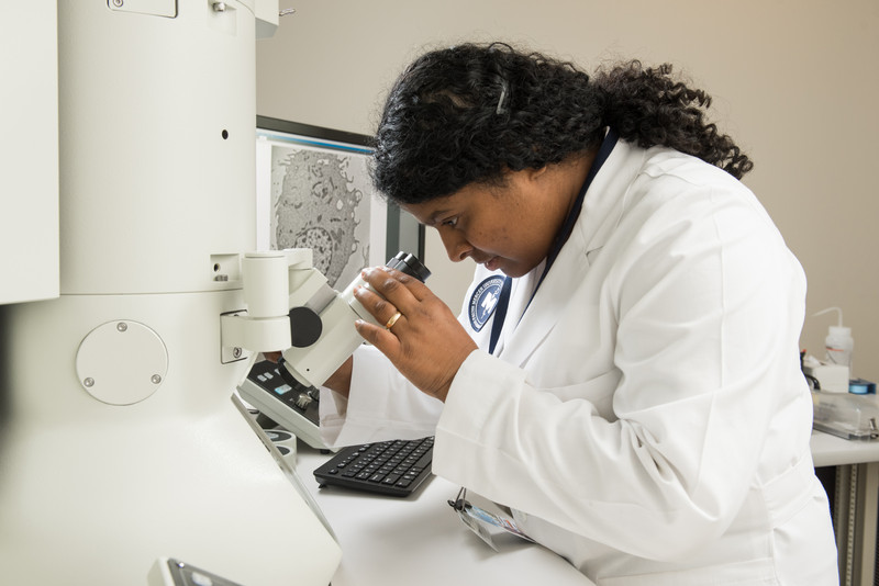
In terms of power and resolution, Mercer’s TEM is tops in southeast Georgia, and in January the university added an important element by hiring molecular biologist Maheshinie Rajapaksha to head the core facility. Dr. Rajapaksha’s arrival opened the door to attracting researchers from other universities to Mercer, both to use the technology and collaborate with Mercer scientists.
“University researchers as far away as Florida and Virginia have already spent time in our facilities generating data with the TEM,” Visalli says.
But the greatest benefit will be the acceleration of research at Mercer, he points out. Joanna Thomas, a biomedical engineering faculty member, is exploring materials science at the atomic level. Joseph Keene, a Mercer chemist, is developing state-of-the-art nano-materials.
“And it’s important to note that research projects involving the TEM include opportunities for undergraduate, graduate and medical students to get involved,” Visalli says.
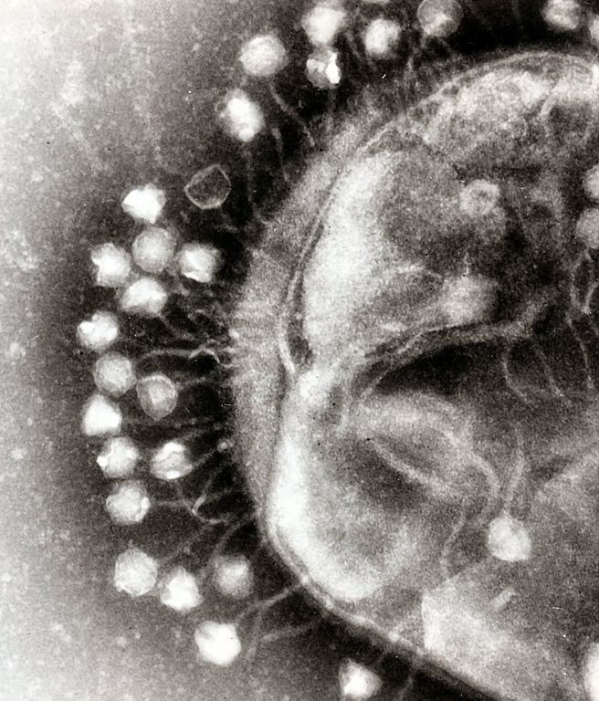
Above: Mercer's Maheshinie Rajapaksha, who oversees the Transmission Electronic Microscope, examines an epithelial cell infected with a virus (depicted on the screen).