Featured Discovery


-
-
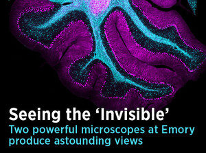
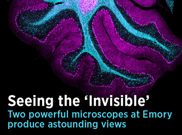
Two microscopes that GRA helped purchase for Emory University are pushing the boundaries of scientific discovery. These powerful instruments perfectly illustrate how investing in a crucial piece of technology helps generate millions in new research funding — and yields new insights into problems facing humankind. Picture: A view inside the cerebellum of a mouse.
-

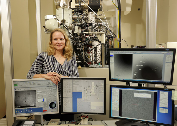
Images of stunning resolution and clarity are the product of Emory’s cryo-transmission electron microscope. Because the microscope uses samples that are frozen — instead of chemically fixed — it gives scientists a better look at the structure of molecules in their natural state. Dr. Elizabeth R. Wright, head of the university’s electron microscopy core, purchased the $1.5 million microscope with funds from GRA and the National Science Foundation.
-
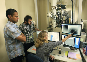
Post-doctoral fellow Dr. Ricardo C. Guerrero-Ferreira loads a bacterial sample into a cryo-holder, which fits inside the microscope for viewing. The bacterial sample is frozen to liquid nitrogen temperatures — around -350° F. The freezing technique keeps the specimen very close to its natural state, unlike the chemical fixatives or heavy metal stains commonly used in microscopy, which can alter the structure of cells. Researchers viewing frozen, unfixed, unstained samples gain a better understanding of exactly how cells and macromolecules are structured.
-

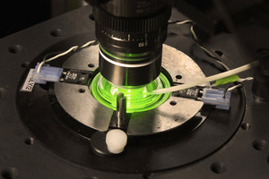
Researchers must work carefully but quickly, so that the samples and equipment are kept extremely cold. If the temperature is low enough, perfectly transparent ice, resembling glass, will form around the sample to bring an unobstructed view of the macromolecules. However, if the sample temperature rises to -150° C or higher, ice crystals can develop, making the sample unusable for imaging.
-
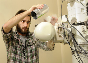
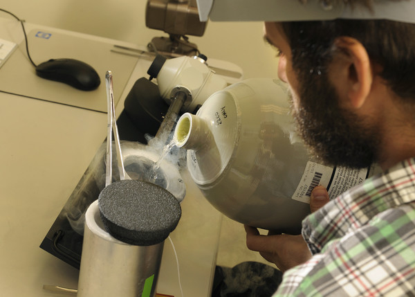
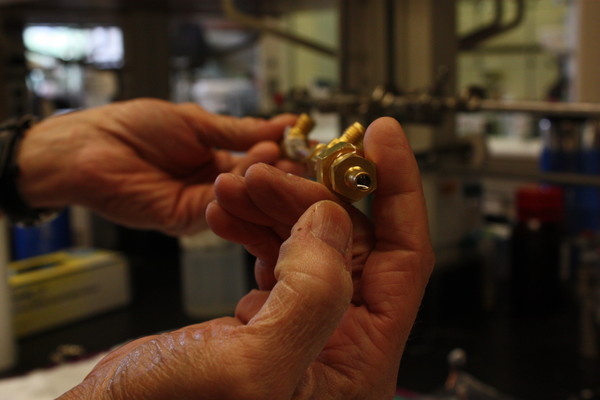
Post-doctoral fellow Dr. Joshua D. Strauss refreshes the liquid nitrogen in the cryo-holder — now inside the microscope — to keep the temperature of the specimen frigid. Using controls, scientists can tilt the bacterial specimen to capture views from different angles. This process, known as electron tomography, can render a three-dimensional model of the macromolecules. The Emory group needs these models to analyze the viruses and bacteria they study, which take asymmetrical, unique shapes and structures.
-
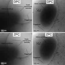
These images show how much added detail is captured by Emory’s cryo-TEM microscope, thanks to its Zernike-style phase plate system. In each pair of images, the top photo was taken without use of a phase plate; the bottom photo shows the improved contrast and resolution with the phase plate. While phase contrast has enhanced light-based microscopy for decades, this technology was only recently developed for electron microscopes. Installations are rare: Emory’s is one of only three 200 kV cryo-TEMs in the United States that has this technology.
-
Wright and her team study images of Vibrio vulnificus, a bacterium that lives in warm seawater. People may become infected with the strain after eating raw oysters or other shellfish. For people with weakened immune systems, V. vulnificus infections can be fatal. Pictures of the bacteria taken with the cryo-TEM have revealed new clues about how they release outer membrane vesicles — small “cargo vessels” containing cytotoxins that weaken host cells, allowing the infection to spread. Wright anticipates her team’s research will help develop antimicrobials that can stop the process.
-
GRA also invested in a multiphoton microscope at Emory to advance research into Alzheimer’s Disease and other brain disorders. The scope has no pinhole aperture; rather, it applies a focused laser beam to excite chemical compounds in a specimen, so that they emit light.
-

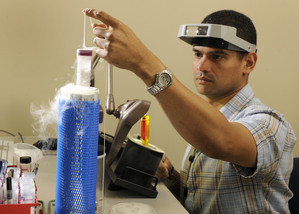
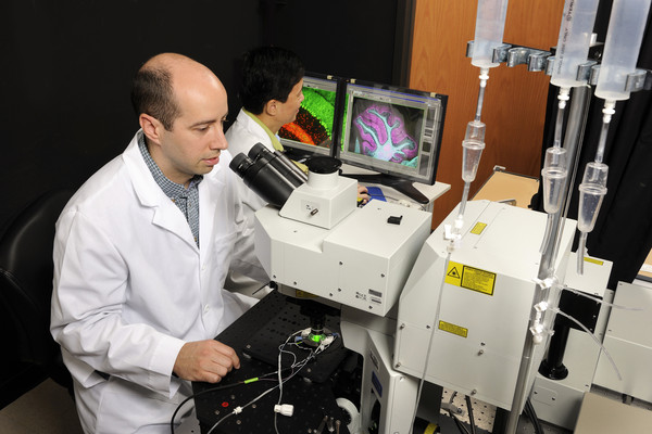
The multiphoton scope enables scientists in the lab of Dr. James Zheng to peer much deeper into brain tissue than other instruments allow. The technique minimizes damage to the tissue, allowing researchers to conduct studies over longer periods of time. Here, Dr. Ken Myers uses the optical viewer to prepare a specimen to be studied. For the microscope to work, the room’s lights are turned off (with the scope shrouded by a black curtain), and the specimen is viewed on a computer monitor.
-
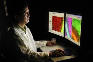
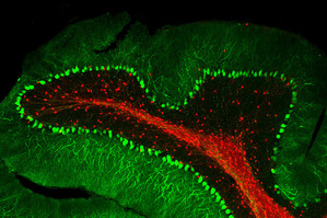
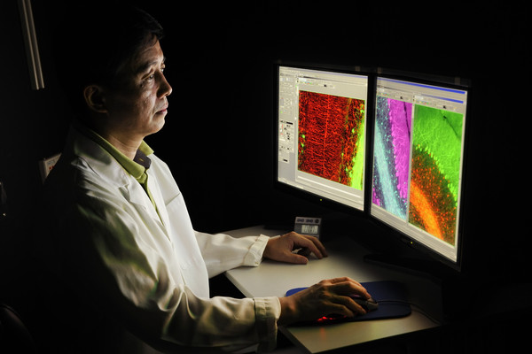
In darkness, Dr. Zheng examines cells and neurons within the cerebellum of a mouse model — images generated by the multiphoton scope. The tissue here was taken post-mortem, but even the brain of a live mouse can be studied using a cranial “window” on top of the skull.
-
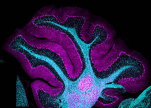
Another view of the cerebellum of a mouse. The combination of chemicals and laser light illuminate areas of the brain with remarkable clarity.


Mouse-over each image for details about GRA's investment in high-powered microscopes.
• Photos by Jack Kearse, Emory University
It's filtration. At the molecular level. And its implications are huge. GRA Eminent Scholar Bill Koros explains why.
GRA's investments in technology give scientists the tools to attract major research funding -- and make surprising discoveries.