Microvesicle Core
Morehouse School of Medicine
Contact: Dr. Ming Huang, MD
Phone: 404-752-1861
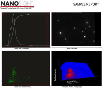
Microvesicles include both microparticles (100nm-1μm diameter) and exosomes (30 to 150 nm diameter). These vesicles are secreted by most cell types and contain a variety of molecules including peptides, mRNA, and microRNA. The Microvesicle Core uses NTA to analyze vesicles. NanoSight NTA instruments generate videos of a population of nanoparticles in a liquid, illuminated by laser light. A specially designed and constructed laser illumination device is mounted under a microscope objective. Particles in the liquid sample which pass through the beam path are seen by the instrument as small points of light moving rapidly under Brownian motion. The laser measures both particle/vesicle size and number.
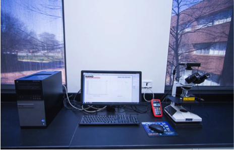
Microvesicle Core Equipment:
Beckman Allegra X-15R Clinical Centrifuge
Beckman Optima TLX, Optima Max-XP, Optima XPN100 Ultracentrifuges
NanoSight LM10-HSBF Nanoparticle Characterization System and NTA 2.3 Analytical Software
Microvesicle Core Services and Data Analysis Services:
1. Isolation and purification of microparticles or exosomes from cell cultures with data analysis.
2. Isolation and purification of microparticle or exosomes from plasma with data analysis.
3. Isolation and purification of microparticle or exosomes from urine with data analysis.
4. Microparticle or exosome data analysis only. If you prefer to isolate exosomes, please resuspend the exosome pellets in 1x PBS and provide >350 µL for NanoSight analysis.
Data Analysis:
1. Microparticle or exosome pictures (including 3D intensity image)
2. Microparticle or exosome video (10 seconds/sample)
3. Microparticle or exosome number determination (particle/mL)
4. Microparticle size (100nm-1µm) or exosomes size (30nm-150nm)
Learn more about research at Morehouse School of Medicine »
< Back to GRA Core Exchange facilities