Center for Systems Imaging Core
Emory University
Contact: John Oshinski, Ph.D., Core Director
Phone: 404-727-5894
The Center for Systems Imaging Core (CSIC) is an Emory University School of Medicine core lab dedicated to providing state-of-the art human and pre-clinical imaging, as well as radiopharmaceutical development to the Emory community. CSIC is the cross-disciplinary scientific, administrative, and educational home for imaging sciences at Emory University. The goals of this center are to: (1) support the advancement of scientific research focused on the development of imaging biomarkers, (2) promote the development and application of biomedical imaging technology particularly magnetic resonance imaging, (3) provide core services for human and animal imaging studies, and (4) build cross-cutting educational and training programs.
The CSI Core is housed in approximately 22,600 square feet across the Emory campus. This total includes a 18,700 square foot facility (G1 10,950, G2 RP 4230, G2 MR 3520) in the Health Sciences Research Building, 800 square feet in Emory University Hospital (EUH), 400 square feet in the Whitehead Biomedical Research Building (WBRB), 2,000 square feet in the Brain Health Center at Emory ‘s Executive Park Campus Building 12 (EP12), and 700 square feet of shared clinical/research space at The Emory Clinic building C (TEC). The director of CSIC is John Oshinski, PhD (jnoshin@emory.edu) and the Medical Director is Ranliang (Ron) Hu, MD, PhD (ranliang.hu@emory.edu). Program Directors are Shella Keilholtz, PhD (pre-clinical MRI), Deqiang Qiu, PhD (MRI), Steven Liang, PhD (PET and Radiochemistry). There are 11 staff members including MRI and PET Technologists, Radiopharmacists, and scientists to provide computer, MRI physics, and small animal support services.
If you use the CSIC for your research, please acknowledge the CSIC in your publications and include its RRID (RRID:SCR_023522).
Major imaging equipment housed at the Health Science Research Building II (HRSB II):
- General Electric Cyclotron
- Radiopharmacy and radiochemistry lab featuring Von Gahlen Hot Cells
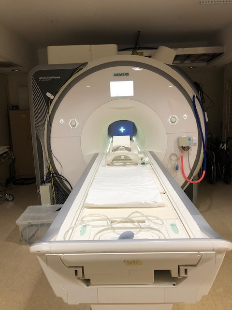
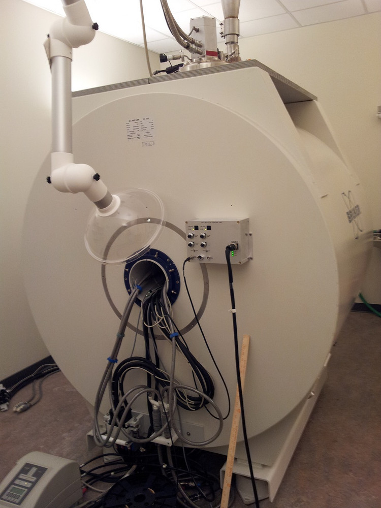
- 3.0 and 7.0T Siemens human MRI systems (PrismaFit and Terra respectively)
- Siemens human PET/CT Biograph Vision 600 system
- Pre-clinical Molecubes B-X Cube micro PET-CT system
- 11.7T Bruker pre-clinical MRI system
- Procedure and testing rooms, changing and consent rooms
- Mock MRI scanner
Major imaging equipment housed at Emory University Hospital (EUH):
- 3.0T Siemens PrismaFit MRI scanner. The presence of this scanner in the hospital allows for in-patient and contrast-enhanced research studies.
Major imaging equipment housed at The Brain Health Center (BHC):
- 3.0T Siemens Prisma MRI scanner
- Image processing and phantom construction lab
Major equipment at The Emory Clinic (TEC-Bldg C):
- General Electric PET/MR scanner
Major equipment at The Whitehead Building (WBRB):
• Bruker 9.4T pre-clinical MRI scanner
Magnetic Resonance Imaging (MRI)
CSIC operates three full-time research dedicated Siemens Magnetom Prisma 3.0T MR scanners and one 7.0T Siemens Terra scanner. With multiple human research 3T MRI scanners, MRI studies can be effectively distributed across the Emory community. Studies that require IV contrast, proximity to Emory University Hospital (EUH) or studies performed on in-patients can be performed on the scanner located on the ground floor of the hospital (CSI-EUH). Outpatient studies, where convenience of parking and more flexible scheduling is required can be performed at scanners located at the HRSB II location. Studies that are performed on subjects seen in the Brain Health Center (BHC) can be enrolled for research studies at that location (CSI-BHC). All three scanners are on the Prisma platform with VE11C software.
3T MRI Scanners. Magnetom Prisma whole-body MR systems are equipped with a state-of-the art gradient system with a maximum per axis strength of 80 mT/m and slew rate of 200 T/m/sec. The system has 64 independent RF receiver channels capable of 204 receiver connections, and a 2-channel RF transmitter. Multiple coils are available, including a 64-channel head/neck coil with 52 channels for imaging of the head region and the remainder for neck imaging, a 32-channel head-only coil, a 20-channel head/neck coil, spine array coil, flexible chest coil, large and small flexible coil for extremity imaging, carotid coil, Tx/Rx CP Head Coil for large no-cap head space, and a 31P dual-tune flexible coil shared across sites for phosphorus spectroscopy. The scanners are equipped with DirectRF and DirectConnect technology, providing a significant increase in signal-to-noise ratio. The Prisma scanner platform allows efficient acquisition of high-resolution fMRI and DTI images with protocols compatible to those released by the Human Connectome Project. Furthermore, the Prisma scanners located at EUH and BHC are equipped with multi-nuclei spectroscopy and additional shimming power for improved magnetic resonance spectroscopy.
A number of advanced research sequences are also available, including vessel wall imaging, quantitative Arterial Spin Labeling, Diffusion Spectrum Imaging (for High Angular Resolution Diffusion Imaging), Simultaneous Multi-Slice EPI (allowing for sub-second high-resolution whole-brain fMRI data acquisition), 4D phase contrast MR for measuring time-resolved flow velocity, displacement encoding with stimulated echoes (DENSE), and multi-echo and ultra-short echo time sequences. Through our master research agreement, advanced work-in-progress MR sequences from the vendor, collaborators from other institutions are available. In addition, locally developed sequences are available.
7.0T MRI Scanner at HRSB II. The Ultrahigh field human MRI scanner is a Siemens Terra 7T with a bore size of 60 cm and a magnet length of 270 cm. It is equipped with Zero Helium boil-off technology as well as passive and active shimming with 3 linear channels (1st order) and 5 nonlinear channels (2nd order) guaranteeing homogeneity of 0.02 ppm over a 20 cm volume. The system is equipped with XR Gradients (80 mT/m @ 200 T/m/s, 400 msec rise time) and 64 channel TIM RF coil technology. The Maximum effective vector gradient amplitude is at 139 mT/m with a maximum slew rate of 346 T/m/s and a duty cycle of 100%. This allows slice thickness (3D) as low as 0.05 mm, and in-plane resolution at 7 µm with maximum matrix size of 1024. The system is equipped with multinuclear imaging and spectroscopy capabilities, and has state-of-the art parallel transmit (PTX) capacity for uniform B1 transmit profile. The system has 1Tx32 R x head coil and a1Tx28Rx Knee Coil for single transmit mode and a 8 channel PTX coil with intergraded 32 channel receive-coil array.
The system operates on Syngo MR11 software with secure Dual Mode functionality for secure switching between research operation (parallel transmit) and clinical operation (FDA approved single transmit mode). The system is equipped with the Neuro and Ortho software applications suites. The system has Wireless Vector ECG, Respiration, and pulse sensors for physiologically synchronized imaging. Application packages include DTI, Resolve, SWI, SMS, and 3D Pace, among others.
Stimulus and response system for functional MRI. All scanners are equipped with peripheral systems for fMRI. Stimulus/response controls for behavioral tasks concurrent with fMRI are supported by an array of hardware specifically designed to allow investigator flexibility and precision. Visual presentation at the EUH site is provided by a high-resolution LCD projection system (1400x1050 SXGA, 4200 lumens, 1300:1 ratio) delivered from the back of the suite onto a custom fit screen mounted within the bore behind the participant’s head. The BHC and HRSB sites are equipped with Cambridge Research Systems BOLDScreen MRI compatible LED displays. Audio presentation is provided by an Avotec Silent Scan 3100 that has been calibrated to maintain sound pressure levels that are dependent directly on input (flat frequency response +/- 4dB, 200-4500Hz range). A fiber-optic ergonomic bilateral button response system from Psychology Software Tools exists, as well as a control unit to support custom response shapes (joysticks, steering wheels, wands) from Current Designs. All of the hardware is connected through a single switch that signals TTL trigger pulses and allows connectivity to an investigator’s laptop with non-proprietary connections (USB, 1/8” minijack audio, VGA & DVI). A dedicated stimulus and response monitoring computer running Eprime 2.0 and Presentation stimulus programming software also exists. An OptoAcoustics FOMRI MRI-compatible microphone featuring advanced active noise cancellation technology is available for speech fMRI paradigms. CSI-EUH MR scanner is also equipped with multi-nuclei option. Currently, a 31P/1H dual tune flexible coil is equipped and is applicable to phosphor MRS and metabolism studies.
HRSB II MRI: Other Equipment. A Biopac MP150 (Goleta, CA) MRI-compatible physiological response measurement system is available for collecting peripheral physiological measures. The MP150 system provides high resolution (16 bit), variable sample rates for analog and calculation channels, 16 analog inputs and two analog outputs, digital I/O lines (automatically control other TTL level equipment), and 16 online calculation channels. The MP150 System provides high-speed acquisition (400 kHz aggregate) via an Ethernet connection to a host computer. AcqKnowledge, the Biopac control and analysis software package is used to control the acquisition and can be used for data analysis. Available physiological measures are cardiac pulse, heart rate, heart period, respiratory sinus arrhythmia, respiration and electrodermal activity. The physiological measurements recorded by Biopac can be viewed real-time on a dedicated laptop computer through the Biopac data acquisition software. All responses can be recorded in MatlabTM, text or proprietary Biopac software formats, for retrospective analysis. A Resoundant™ MR Elastrography (MRE)system is available on the scanner and is integrated into the acquisition trigger system. Both head and liver drivers are available.
EUH MRI: Other Equipment. Peripheral equipment, including computers and software for paradigm generation, set up for stimulus presentation, devices for recording behavioral data and physiological parameters including heartbeat, respiration, blood pressure, eye movement, ECG, EEG, and EMG are also established for operation concurrent with MR acquisition. Stimulus generation and presentation setup allows us to present acoustic, electric, and vibrational stimuli and oral and venous administration of liquids. Setup for response via either button box, keyboard, mouse, speech, eye movement, and grip force have been established. We are also equipped with an electronic shop and a small machine shop, providing the capability to fabricate custom MRI coils, animal holders, and special purpose stimulation devices. Other equipment in the scanner rooms includes an Ohmeda Biox 3700 Pulse oximeter, a Sage 351 infusion/withdrawal syringe pump, and a Dinamap 1846 SX Critikon vital signs monitor. A MEDRAD (Warrendale, PA) Power Injector system for contrast administration is available.
BHC MRI: Other Equipment. A Biopac MP150 (Goleta, CA) MRI-compatible physiological response measurement system is equipped to drive oxygen and carbon dioxide concentration of exhale air flow. The MP150 System provides high-speed acquisition (400 kHz aggregate) via an Ethernet connection to a host computer and is ready to integrate other BIOPAC peripheral physiological measuring modules. AcqKnowledge, the Biopac control and analysis software package is used to control the acquisition and can be used for data analysis. A three gas bottles gas distribution system is wired to provide inhaling gases with different oxygen and carbon dioxide concentrations to the research participant. The data recorded by Biopac can be viewed real-time on a dedicated desktop computer through the Biopac data acquisition software. All responses can be recorded in MatlabTM, text or proprietary Biopac software formats, for retrospective analysis. An MRC eye-tracking system is equipped to monitor and record eye motion data.
Mock MRI Scanner at HRSB II. A mock MR scanner is set up in the HRSB lI location. This mock scanner is similar in appearance to the Siemens MRI scanners. It provides stimulus presentation and scanner noise emulations and is used to familiarize pediatric subjects with MRI scanner operation and to acclimate them to the MR scanning environment.
Cyclotron, Radiochemistry, and Radiopharmacy at HRSBII
Cyclotron and Radioisotope Production at HRSB II. The Cyclotron Suite houses a General Electric PETrace 880 18MeV self-shield cyclotron. This cyclotron allows the production of Fluorine-18, Carbon-11 (carbon dioxide), Nitrogen-13 (ammonia), Oxygen-15, and Gallium-68. The cyclotron includes the PROCAB processing system for Carbon-11 and a processing system for conversion of Oxygen-15 into [O-15] Water. The high energy cyclotron allows for single bombardment production of energies up to 100uA or dual bombardment up to 130uA . The cyclotron also has the capability of Deuteron beam at 8.4MeV for production of Oygen-15 without enriched target material. The transfer of radioisotopes from the cyclotron to the Radiopharmacy and Radiochemistry Labs is via a Von Gahlen Active Distribution System providing the delivery of isotopes to a variety of locations within the labs.
Radiopharmacy and Human Tracer Research Development at HRSB II. The CSIC PET Radiopharmacy is a Georgia State-Licensed Nuclear Pharmacy directed by Dr. Steven Liang, PhD, and managed by Ronald Crowe, RPh, BCNP, a licensed Radiopharmacist. The Radiopharmacy facility is operated under the guidelines outlined in Unites States Pharmacopeia chapters <823> and <825> and includes space for research radiopharmaceutical production, and ISO7 certified cleanroom and dispensing spaces. The Production space utilizes Von Gahlen cGMP Stacked Synthesis Hot Cells with adjacent cabinets for preparatory-HPLC equipment as necessary for radiopharmaceutical production. The Synthesis Hotcells contain the following GE Radiochemistry production systems: TRACERlab FX2 N, TRACERlab FX2 C, Fastlab 2 and Fastlab 2 with the Developer System and HPLC+ Purification and Reformulation Systems. Additionally, the Synthesis Hot Cells house a Trasis All-In-One AIO36 Radiochemistry production system. The production area also houses the following systems for quality assurance testing: Agilent HPLC with RAM detection, Agilent Gas Chromatography with RAM detection, LabLogic Scan-RAM TLC, LabLogic Bubble Point Tester, Charles River Nexgen PTS BET testing unit, and Sterility Incubation cabinets. A SwissLog Pneumatic System provides the shielded transport of radiopharmaceutical doses to various scanning locations throughout HSRB2. The space is monitored by the nuviaTECH ALMO radiation detection systems to monitor and alert for radiation levels.
The ISO7 Cleanroom space houses a Baker EdgeGARD laminar flow hood providing an ISO5 certified space for critical manipulations not involving radioactive isotopes. The ISO7 Dispensing space utilizes Von Galen cGMP Modular Hot Cells with laminar cross flow for the dedicated handling and dispensing of F-18 and C-11 radiopharmaceuticals, respectively. The Modular Hotcells are equipped with Central Research Labs Model G-LD Remote Manipulators. The space also contains a Von Gahlen cGMP Shielded Glove Box for handling and dispensing Ga-68. All three contain Capintec CRC55tPET dose calibrators for accurate measurement of radiopharmaceuticals. The space is monitored by the nuviaTECH ALMO radiation detection systems to monitor and alert for radiation levels.
The Radiopharmacy also contains separate spaces for secure materials acceptance and control, glassware depyrogenation process oven, refrigeration and -20ºC materials storage units, and a Mirion Shielded Multi-Channel Analyzer. Additionally, the Radiopharmacy can provide delivery of Radiopharmaceuticals to imaging locations on the Emory Clifton Campus utilizing Biodex PET Vial Pig Shipping Systems via study coordinated courier service.
Positron Emission Tomography and Computed Tomography (PET/CT) System at HRSBII
The PET/CT system is a fully integrated Siemens Biograph Vision 600 system with a 78 CM bore and 136 cm tunnel length. The PET system features 3.2-mm lutetium oxyorthosilicate (LSO) crystals that are 100% covered by SiPM s2Optiso Ultra Dynamic Range (UDR) detector technology which enable higher sensitivity and lower dose. The detector technology features 48-mm3 volumetric resolution and 214-ps "true" time-of-flight performance. The small detector block size delivers >1789 kilo counts per second effective peak NECR. The High-flow direct-cooling of the detector electronics assembly allows the detector temperature to remain stable at room temperature for outstanding performance and serviceability, as well as improved patient comfort. Software includes: FlowMotion™ AI, Multiparametric PET AI, OncoFreeze™ AI, QualityGuard™, and FAST CARE CT technologies.
PET/MR System: A Shared Research/Clinical resource at TEC
In March 2021, Emory University installed 3 Tesla PET/MR system from General Electric (GE) Healthcare. The system was purchased with funds from an NIH S10 large instrumentation grant and supplemented with Emory Healthcare funds. The scanner is located in a translational area within the Emory Clinic (TEC, across Clifton Road from Emory University Hospital) and operates under as shared use agreement to support both research and clinical use. The CSI Core manages the scanner on research days. The scanner is a GE SIGNA PET/MR 3.0T 26 with Quantworks features a simultaneous time of flight (TOF) PET imaging with whole body 3.0T magnetic resonance imaging (MRI) in a 60 cm bore. The PET system is composed of 45 LBS-Lutetium based scintillator rings (20,160 total crystals) and 28 Silicon Photomultiplier Modules. The timing resolution is <400 psec with a coincidence window of 4.57 ns. Axial field of view is 25.0 cm, the trans-axial field of view is 60 cm. Trans-axial resolution is 3.7-4.2mm and axial resolution is 4.8-7.1mm. The PET processing system contains 896 1.15 GHz GPU cores and a six-core Dual Intel Xeon Processor CPU. The system is equipped with a number of viewing and image processing programs, including zero-TE attenuation correction for the head. The MR system is a 3.0 Tesla GE SIGNA 750W with gradients capable of a Peak Amplitude 44 mT/m and a Peak Slew Rate 200 T/m/s. Main field homogeneity is <0.500 ppm over 40 cm FOV. The system comes equipped with a number of coils including spine coil, body array coils and Transmit/Receive head coils. The software suite includes advanced neuro, body, oncology and orthopedic, cardiovascular, and pediatric packages and includes multi-nuclear spectroscopy.
MicroPET/CT animal Scanner at HRSBII
The pre-clinical Molecubes B-X Cube micro PET-CT system was installed in HSRB-II in November 2024. The system is a combination of PET and CT and is a high-performance innovative benchtop scanner. The state-of-the-art 5-ring 45 detector system ensures best-in-class sensitivity while maintaining uniform sub-millimeter image resolution across the entire field-of-view. The introduction of monolithic scintillators coupled with Peltier controlled SiPM detectors along with their iterative image reconstruction techniques results in superior image quality. The Molecubes microCT is a high-throughput benchtop scanner. It allows for fast whole-body imaging at an extremely low dose and excellent soft tissue contrast. The shelf-shielded system is a truly mobile in-vivo scanner due to its compact design and light weight. Advanced workflows such as gated and dynamic contrast enhanced imaging can be achieved in a functional and integrated setup. The cube system’s iterative reconstruction techniques and expert user mode make for a multi-purpose solution for most pre-clinical imaging applications. This PET/CT combination optimally fits whole body rodent molecular imaging. Intuitive acquisition, reconstruction and viewing software allow for highly efficient multi-modality scanning with unsurpassed image quality. A high-performance workstation supports the imaging workflow - control, display and analysis of large image volumes. The Molecubes software suite, CUBEFLOW, allows for intuitive and efficient workflow to maximize researchers’ accuracy and speed. The software platform integrates quality control, physiological monitoring, image acquisition and advanced reconstruction techniques.
11.7T Bruker Pre-clinical MRI System at HRSB
The ultra-high field preclinical scanner is a BioSpec 117/16 USR multipurpose scanner for magnetic resonance imaging (MRI) and magnetic resonance spectroscopy (MRS). The main field is 11.7 Tesla, with a 16 cm bore USR magnet and a High-Power Gradient Amplifier. The system operates on the ParaVision 360 MR Comprehensive Imaging Workplace. The system is equipped with two 1H parallel transmit and receive RF channels and a Broadband transmit and receive channel supporting the following applications:
- 1H, 19F, and X-nuclei with MR frequencies from 15 MHz up to 31P
- 1H arterial spin labeling (ASL)
- X-nuclei MRI/MRS with 1H decoupling (WALTZ, MLEV)
- MRI/MRS with hyperpolarized nuclei (13C, 129Xe)
The system is equipped with a circularly polarized, transmit/receive MRI RF Coil with a 72 mm ID, as well as an Anatomically shaped four element array coil for mouse brain investigations.
The system is equipped with the MR Imaging Plus Package extends the ParaVision 360 imaging portfolio with the following MRI imaging methods for preclinical research:
- Diffusion imaging methods: DTI-SE, DTI-EPI, T1-EPI, T2-EPI, T2*-EPI, DTI-Spiral
- fMRI, DCE and Perfusion methods: EPI, CASL-EPI, FAIR-EPI, FAIR-RARE Spiral
- IntraGate wireless cardiac imaging methods: IgFLASH, IgUTE
- Advanced Angiography: Flowmap
- Short TE: UTE, UTE3D, ZTE, RAREst
The MR Spectroscopy Package extends the ParaVision 360 imaging portfolio with state-of-theart MR Spectroscopy and Spectroscopic Imaging methods for preclinical research including:
- Localized Spectroscopy: PRESS, STEAM, ISIS
- Chemical Shift Imaging: CSI, EPSI
9.4T Bruker Pre-clinical MRI System at WBRB
A 9.4T/21-cm Bruker animal MR imaging/spectroscopy system is housed in the Whitehead Biomedical Research Building in approximately 400 square feet of space. The magnet is actively shielded to reduce the extent of the fringe field. The imaging console is interfaced with a Bruker AVANCE spectrometer driven by a LINUX workstation and Bruker ParaVision 5.1 imaging software. The system is equipped with actively decoupled RF coils (volume coil as a transmitter and the surface coil as a receiver) with 2-RF channels: one with 1000 Watt RF amplifier for 1H NMR studies and the other with 800 Watt broadband amplifier (frequency range from 6 to 365 MHz) for X-nucleus NMR studies. A quadrature volume coil optimized for imaging rat brains and a variety of surface coils are also available. The scanner is equipped with a state-of-the-art BFG 200/115-S-14 12-cm diameter gradient insert from RRI (maximum gradient strength 675 mT/m, 120 usec risetime), two actively shielded Bruker BGA gradient sets, BGA-12 (12 cm, maximum gradient strength of 400 mT/m, 88 usec risetime) and BG-6 gradient set (6 cm, maximum gradient strength of 1000 mT/m, 55 usec risetime), all driven by Copley 200A/300V Gradient Amplifiers. Peripheral equipment including a) physiological signals monitoring system (BioTrig), used for synchronizing MR acquisitions with ECG or respiration triggering signals; b) animal anesthesia and physiology maintenance system; c) a comprehensive set of RF coils, suitable for studying different sized animals, different tissues and locations, and different nuclei including proton, 17O, 13C, 19F, an 31P; d) acoustic, optic, and electrical stimulation accessories for functional study. This system is suitable for studying mice, rats, ferrets and other small animals.
Computing Facilities
The CSI Core is equipped with a state-of-the art computing facility consisting of a twenty-four nodes linux mini cluster, disk RAIDs with 1569 TiB total storage capacity, a full rack capable of supporting up to 16U calculation nodes, including 1016 CPU threads and 6 NVidia P100/A40 powered GPU nodes, and three automated off-site back-up server with 1145TiB backup storage configured to bi-weekly rotation and one-year time machine backup scheme. For data processing and analysis, Matlab, IDL, LCmodel, SPSS, SPM, fsl, AFNI, freesurfer, ANTs, MrTrix, Slicer, tensorflow, and a variety of python-based pipelines are installed and available on our cluster. VMware and Oracle VirtualBox are also available. The computer cluster is also a dicom receiving server open to all collaborating PIs for data storage and analysis purposes.
More about this core facility at Emory University »
Joy Staulcup (Operations Manager), Phone: 404-712-1024
< Back to GRA Core Exchange facilities