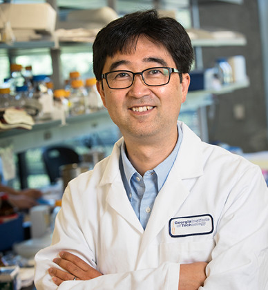
Shuichi Takayama, Ph.D
Micro & Nano Biotechnology
Georgia Institute of Technology
Recruited: 2017
Imagine a computer chip as wide as the eraser of a pencil, etched with channels smaller than a human hair – sometimes thousands of times smaller – flowing with materials too tiny to see. At this scale, liquids behave very differently, which opens new avenues for experimentation. Scientists can do things working with less than a droplet of fluid that they would not be able to do on a larger scale.
This is the research world that Shuichi Takayama inhabits. It’s called microfluidic and nanofluidic technology, and it involves creating and studying miniscule devices for manipulating incredibly small amounts of fluid – all for the purpose of advancing medicine.
A major application for this technology is a so-called “organ-on-a-chip” system. Using a small amount of biological material, researchers can create a “microenvironment” that mimics conditions found inside a kidney, liver or other organ. Unlike a flat and static layer of cells in a petri dish, this microenvironment is three-dimensional and dynamic, which offers a much better representation of how cells actually live and interact.
Takayama is developing many of these groundbreaking organs-on-a-chip devices. One example is a recent breakthrough, the lung-on-a-chip. Takayama and his colleagues are using the device to study the effects of fluid in the lung, a common symptom of lung disease.
Often partnering with clinician experts in health and biomedical research, Takayama and his team are perfecting a handful of similar devices. Their kidney-on-a-chip device is helping researchers measure the best way to administer antibiotics to minimize kidney toxicity. Their miniature cancer models are providing important tools for understanding how cancer cells grow and migrate. Another of their microfluidic devices mimics the chemical stimuli cells use to communicate, shedding light on how cell signaling works in real-time.
Not only do such devices provide tools for studying the behavior of cells and illuminating the mechanics of disease, they also provide a powerful method for testing potential therapeutics more rapidly. Researchers can introduce potential drug compounds to these engineered microenvironments and get fast information on how the drug impacts, say, a diseased heart cell. The technology could help usher in new medicines for many of the complex health conditions that have so far eluded effective treatment.
Takayama’s team has also received wide acclaim for their extensive work with assisted reproductive technology. He and his colleagues have developed microfluidic technologies to advance artificial fertilization, including an embryo culture system and a sperm sorter.
The embryo culture system mimics the microfluidic environment surrounding a brand-new embryo inside a mother’s body, giving an in vitro-fertilized embryo a stronger start in the three to five days before being implanted in the mother’s uterus. The sperm sorter is like a microscale maze or racetrack, helping doctors identify the strongest candidates for artificial insemination. Both technologies have been used successfully in the offices of fertility doctors.
Takayama has begun pushing the nanoscale technology into new frontiers by using it to study epigenetic changes to DNA. Epigenetics refers to how DNA is packaged, and changes in packaging can have drastic effects on gene expression (how DNA is used by each cell). Epigenetic studies require large amounts of DNA and multiple strands, with assessment of epigenetic changes based on inference and probability. Takayama’s team has devised a way to stretch out a single strand of chromatin – the genetic material that forms chromosomes – across a nano-channel, and take a picture of it using high-powered microscopy. This technique allows them to look directly at the packaging and physical properties of DNA, opening the door to an unprecedented study of epigenetics.
Takayama and his team have spent years developing microfluidic and nanofluidic technology and refining miniature models of disease. As the research continues, the team is applying these methods to develop better diagnostics and therapeutics for a whole host of diseases.
Takayama is also exploring how to add an immune-system component to these organ-on-a-chip devices, mimicking not just the behavior of lung cells but the interplay of immune cells. Georgia Tech’s Center for Immunoengineering, launched in 2013, is an excellent asset and resource in this work.
Research
- Microfluidic models of cancer metastasis
- In vitro fertilization on a chip
- Organs-on-a-chip systems
- Aqueous two-phase systems (ATPS)
- Validation of protein biomarkers of disease
- Understanding cell signaling mechanisms
- Microenvironment engineering
- Size-adjustable nano-channels and DNA linearization
Straight from the Scholar
“There’s a lot of great super-resolution imaging expertise right here in our building at Georgia Tech. And there’s the new Marcus Center for cell manufacturing [MC3M], which is great for our cell therapy and cell analysis research. We do like the Emory connection as well. In the end, we really want to impact diseases and patients, so the connection with Emory’s hospitals is really important.”

